Thanks to funding from the VRT Foundation for the valorisation of Trentino research, ProM Facility will contribute to the work carried out by the University of Trento research group that has developed a system to improve the monitoring of the circulatory system. This new technology can have multiple applications, with the aim of making early diagnoses and of monitoring the effectiveness of ongoing therapy, for example in the presence of tumours or chronic diseases such as Alzheimer’s, all pathologies that alter the vascular system.
Imaging of the circulatory system
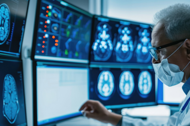
The ultrasound laboratory (Ultra) of the Department of Engineering and Information Science (Disi) at the University of Trento has published a study in the scientific journal ‘Ieee Transactions on Medical Imaging’ that promises to significantly improve the quality of visualisation of the vascular system with an interpolation system of images acquired by means of ultrasound at a low frame rate, or more simply a system capable of accurately reconstructing vascular structures. The current system, in fact, can provide only low quality in depth images or images with very high resolution but at a superficial level.
An ultrasound scanner and a contrast liquid composed of micro-bubbles, each the size of a red blood cell, are used to visualise the circulatory system, in depth and with high image quality, which is useful for early detection of any abnormalities in the shape of blood vessels and for discovering new blood vessels that are created to feed malignant neoplasms. Moreover, the proposed solution uses ultrasound and not radiation and is therefore safe for the patient and repeatable over time.
The first 3D vascular model: the brain
The next stage of this research will be the production of a 3D vascular model, an essential step before human trials. Target areas have already been identified: the brain, the prostate, the breast, the thyroid, because they are easy to reach with clinical scanners and on which later on it will be possible to carry out the first clinical trials together with the Provincial Health Services Agency of the Province of Trento.
The brain will be the first organ realised in 3D by the ProM Facility, which is essential for the development and testing of algorithms dedicated to visualising the vascular network and its properties, in order to validate and test this new imaging technology.
ProM Facility to support Trentino medical research
The 3D realisation of organs to test the HD navigator for the vascular system developed by the University of Trento’s ultrasound laboratory (Ultra) is just the most recent of the Trentino Sviluppo facility’s commitments in the health and medical field.
Worthy of mention also are:
- the production of customised cups for women who have undergone mastectomy – ONEBra
- the development of devices applied to proton therapy using advanced 3D printing techniques – Proton3D
- the reproduction of an aorta model, faithful to the original, of a patient on the list for urgent surgery – aorta in 3D